
Mechanical ventilation is one of the most frequently used life-support interventions in the Pediatric Intensive Care Unit (PICU). Yet, one of the most common mistakes among junior residents is focusing on ventilator settings rather than the patient’s physiology. Effective pediatric ventilator monitoring is what separates experienced intensivists from beginners, since successful ventilation depends less on changing ventilator parameters and more on understanding what the monitored variables are telling us about lung mechanics, gas exchange, and disease progression.
Modern ventilator management is therefore a continuous cycle of assessment, interpretation, intervention, and reassessment. Every number on the ventilator should answer a clinical question. Is oxygenation improving? Is compliance worsening? Is the child developing airway obstruction? Is this patient ready to wean? Consistent pediatric ventilator monitoring at the bedside — rather than relying on isolated arterial blood gas results — is what allows clinicians to catch these changes early and adjust care before a small problem becomes a crisis.
Step 1. Begin with the Patient - not the Ventilator
Before interpreting any ventilator parameter, perform a rapid bedside assessment.
Evaluate:
- Airway position and endotracheal tube depth
- Chest expansion symmetry
- Breath sounds
- Work of breathing
- Patient-ventilator synchrony
- Sedation and comfort
- Hemodynamic status
- Pulse oximetry trend
- End-tidal CO₂ waveform
A falling oxygen saturation may indicate worsening lung disease, but it may also result from accidental extubation, mucus plugging, pneumothorax, low cardiac output, or equipment malfunction. Never treat numbers before examining the Patient.
Step 2. Assess Oxygenation Systematically
Oxygenation reflects the interaction between lung recruitment, ventilation-perfusion matching, and cardiac function. Three indices should become routine tools for every PICU resident.
For quick-reference formulas used throughout this guide, see the PediRex respiratory formulas page.
1. PaO₂/FiO₂ Ratio (P/F Ratio)
Formula: P/F = PaO₂ ÷ FiO₂
Interpretation:
- 300 = Normal
- 200–300 = Mild oxygenation impairment
- 100–200 = Moderate
- <100 = Severe respiratory failure
The P/F ratio is simple and rapidly identifies worsening gas exchange but ignores ventilator pressures. Consequently, two patients with identical P/F ratios may require vastly different ventilator support.
2. Oxygenation Index (OI)
Formula: OI = (FiO₂ × Mean Airway Pressure ×100) ÷ PaO₂
OI incorporates both oxygen requirement and ventilator intensity, making it a superior marker of disease severity.
General interpretation:
- <5 = Mild disease
- 5–10 = Moderate
- 10–16 = Significant impairment
- 16 = Severe respiratory failure
- 25–30 = Consider rescue therapies
- 40 = Consider ECMO in appropriate candidates
OI is the preferred oxygenation metric in severe pediatric ARDS because increasing MAP may maintain PaO₂ despite worsening lung injury.
3. Oxygen Saturation Index (OSI)
When arterial blood gases are unavailable: OSI = (FiO₂ × MAP ×100) ÷ SpO₂
OSI provides a reliable non-invasive estimate of oxygenation severity and is increasingly incorporated into pediatric ARDS assessment. Residents should calculate OSI every few hours in unstable patients to monitor response to recruitment maneuvers, prone positioning, or PEEP adjustments.
Clinical Scenario 1
A 4-year-old with severe pneumonia is ventilated on: FiO₂ 0.80, MAP 16 cmH₂O, PaO₂ 58 mmHg, P/F ratio = 72.
OI = 22
Although the PaO₂ appears acceptable after increasing ventilator pressures, the OI demonstrates severe oxygenation failure. This child requires immediate reassessment for lung recruitment, prone positioning, optimization of PEEP, inhaled pulmonary vasodilators (where appropriate), and discussion of extracorporeal support if deterioration continues.
Step 3. Monitor Respiratory Mechanics
Understanding respiratory mechanics allows clinicians to determine why oxygenation or ventilation is deteriorating.
Dynamic Compliance
Dynamic compliance reflects both lung elasticity and airway resistance. Low compliance suggests:
- ARDS
- Pulmonary edema
- Atelectasis
- Pneumonia
- Pleural disease
Progressively falling compliance often precedes worsening oxygenation.
Static Compliance
Static compliance removes the influence of airway resistance and better reflects true lung stiffness. A declining static compliance despite stable airway pressures usually indicates progression of restrictive lung disease.
Airway Resistance
Airway resistance increases with:
- Bronchospasm
- Bronchiolitis
- Secretions
- Kinked endotracheal tube
- Small endotracheal tube
When peak inspiratory pressure rises while plateau pressure remains unchanged, think airway resistance before increasing ventilator pressures.
Time Constant
Time Constant = Resistance × Compliance
It predicts how rapidly lungs fill and empty.
Short time constants:
- ARDS
- Neonatal respiratory distress syndrome
Long time constants:
- Asthma
- Bronchiolitis
- COPD
Patients with prolonged time constants require longer expiratory times to prevent air trapping and auto-PEEP.
Clinical Scenario 2
A ventilated infant with RSV bronchiolitis suddenly develops: Rising PIP, Stable plateau pressure, increasing ETCO₂ and Wheezing.
Diagnosis: Increased airway resistance.
Management: Suction, Bronchodilator therapy, Longer expiratory time, Reduce respiratory rate, Exclude mucus plugging.
Increasing PEEP alone would worsen dynamic hyperinflation.
Step 4. Evaluate Gas Exchange Beyond Oxygen Saturation
Pulse oximetry provides only part of the picture.
Advanced monitoring includes:
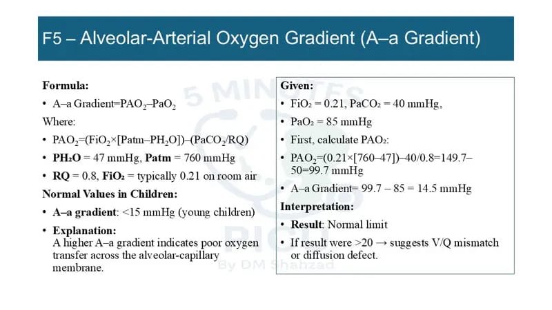
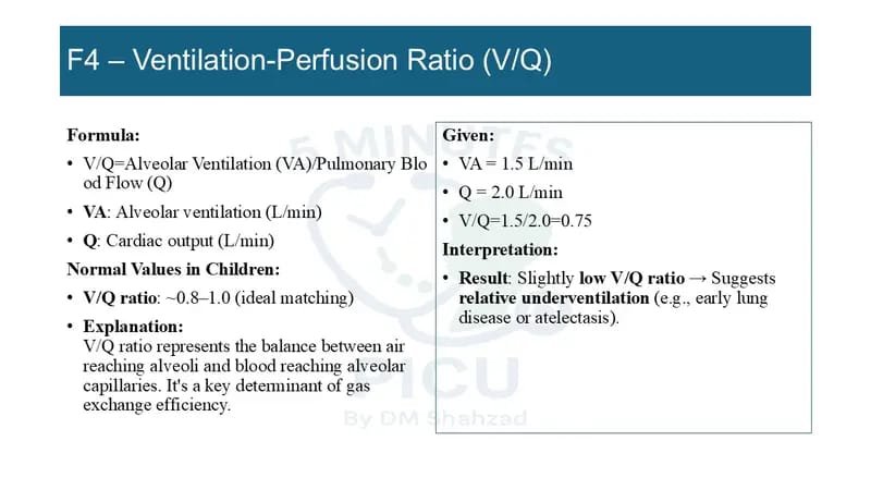
A–a Oxygen Gradient
A rising A–a gradient suggests worsening diffusion impairment, shunt, or V/Q mismatch despite apparently acceptable oxygen saturation.
Physiologic Dead Space (Vd/Vt)
Elevated dead-space fraction indicates ineffective ventilation and is associated with increased mortality in pediatric ARDS. Common causes include:
- Pulmonary embolism
- Overdistended alveoli
- Severe ARDS
- Low pulmonary blood flow
A rising PaCO₂ despite stable minute ventilation should prompt consideration of increasing dead space rather than simply increasing respiratory rate.
Shunt Fraction
Persistent hypoxemia despite FiO₂ of 1.0 usually reflects true intrapulmonary shunt rather than hypoventilation. Management should focus on alveolar recruitment – not merely increasing FiO₂.
Step 5. Advanced Indices Every Fellow Should Know
Mean Airway Pressure (MAP)
MAP is the principal determinant of oxygenation. Increasing MAP improves alveolar recruitment but also increases:
- Barotrauma
- Volutrauma
- Pulmonary vascular resistance
- Right ventricular afterload
Every increase in MAP should therefore be balanced against cardiovascular effects.
Driving Pressure
Driving Pressure = Plateau Pressure − PEEP
Although pediatric evidence is evolving, increasing driving pressure correlates with ventilator-induced lung injury. Maintaining the lowest effective driving pressure while ensuring adequate ventilation is an important component of lung-protective ventilation.
Mechanical Power
Mechanical power integrates:
- Respiratory rate
- Tidal volume
- Airway pressure
- Flow
Rather than focusing on individual ventilator settings, it estimates the total mechanical energy delivered to the lungs. Excessive mechanical power increases the risk of ventilator-induced lung injury, particularly during prolonged ventilation.
Clinical Scenario 3
A child with severe ARDS has:
- Stable P/F ratio
- Rising OI
- Falling compliance
- Increasing driving pressure
- Increasing mechanical power
Despite an apparently stable oxygen saturation, lung injury is progressing.
Appropriate management includes reassessing tidal volume, limiting plateau pressure, optimizing PEEP, considering prone ventilation, evaluating fluid balance, and discussing rescue therapies rather than simply increasing FiO₂.
A Practical Bedside Monitoring Checklist
During every PICU round, ask six questions:
- Is oxygenation improving? (P/F, OI, OSI)
- Is ventilation adequate? (PaCO₂, ETCO₂, minute ventilation)
- Are respiratory mechanics improving? (Compliance, resistance)
- Is lung injury increasing? (Driving pressure, plateau pressure, mechanical power)
- Is the Patient synchronizing with the Ventilator?
- Is the child improving enough to begin weaning?
These questions transform routine ventilator checks into meaningful clinical decision-making.
Key Clinical Pearls
- Never adjust ventilator settings based on SpO₂ alone.
- Oxygenation indices are trend markers, not isolated numbers.
- OI is superior to the P/F ratio in severe pediatric respiratory failure because it incorporates ventilator intensity.
- OSI is an excellent non-invasive alternative when arterial blood gases are unavailable.
- Rising airway pressures should always be separated into increased resistance versus decreased compliance.
- Improving oxygenation at the expense of excessive ventilator pressures may worsen ventilator-induced lung injury.
- The best ventilator strategy is one that achieves adequate gas exchange while minimizing lung injury.
References
Khemani RG, Smith LS, Zimmerman JJ, Erickson S. Pediatric Acute Respiratory Distress Syndrome: Definition, Incidence, and Epidemiology.
Khemani RG, et al. Pediatric Acute Respiratory Distress Syndrome: Consensus Recommendations From the Second Pediatric Acute Lung Injury Consensus Conference (PALICC-2).
Newth CJL, Venkataraman ST, Willson DF, et al. Mechanical ventilation support in children.
Slutsky AS, Ranieri VM. Ventilator-induced lung injury. N Engl J Med.
Tobin MJ. Principles and Practice of Mechanical Ventilation. 4th ed.
Fuhrman BP, Zimmerman JJ. Pediatric Critical Care. 6th ed.
Wheeler DS, Wong HR, Shanley TP. Pediatric