


A 2-day-old term neonate, birth weight 3.2 kg, is admitted urgently to the Pediatric Cardiac ICU for profound cyanosis noted shortly after birth. Pregnancy and delivery were uncomplicated, and Apgar scores were reassuring. However, within hours, the infant developed persistent hypoxemia with oxygen saturations in the 60s despite 100% oxygen.
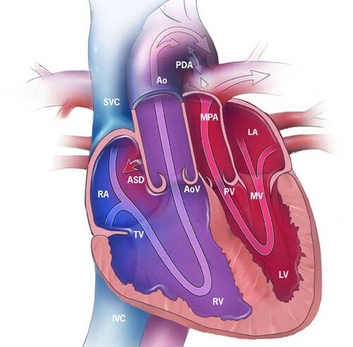
The infant is tachypneic but not in severe respiratory distress. Blood pressure is stable, pulses are palpable, and there is no obvious murmur. A diagrammatic cardiac image (shown above) is reviewed during assessment.
Image-Focused Question
• What abnormal circulatory pattern is demonstrated in this image, and why does it lead to severe cyanosis in the immediate newborn period?
Progressive Clues (Reveal Sequentially)
• Clue 1: The systemic and pulmonary circulations appear to run in parallel rather than in series.
• Clue 2: Oxygenated blood repeatedly cycles through the lungs without reaching the body.
• Clue 3: The infant’s oxygen saturation improves only transiently with maneuvers that enhance atrial mixing.
Answer & Explanation
Key Findings in the Image
• The image depicts discordant ventriculoarterial connections, where:
• The aorta arises from the right ventricle
• The main pulmonary artery arises from the left ventricle
• The systemic (blue) and pulmonary (red) circulations run in parallel
• Inter-circulatory connections (ASD, VSD, PDA) are the only routes for oxygenated blood to reach systemic circulation
Pathophysiology Explained (ICU-Relevant)
• This anatomy results in complete separation of oxygenated and deoxygenated blood:
• Deoxygenated systemic venous blood returns to the right heart → pumped back to the body via the aorta
• Oxygenated pulmonary venous blood returns to the left heart → pumped back to the lungs
• Life depends on mixing at the atrial, ventricular, or ductal level
In the early neonatal period:
• Closure of the ductus arteriosus or restriction of the atrial septum leads to sudden cardiovascular collapse
• Supplemental oxygen alone is ineffective because the problem is circulatory, not pulmonary
Diagnosis (Neonatal Cardiac ICU)
• d-Transposition of the Great Arteries
• Parallel circulations, profound early cyanosis
• Minimal murmur, clear lungs
Management Pearls (Cardiac ICU Focus)
Immediate priorities:
• Maintain ductal patency to allow mixing
• Promote atrial-level communication if restrictive
• Avoid delays in definitive anatomical correction
Monitoring & physiology:
• Pre- and post-ductal saturations
• Lactate and acid–base status
• Echocardiographic assessment of atrial septal flow
Ventilation considerations:
• Avoid excessive oxygen or alkalosis that may reduce pulmonary vascular resistance too rapidly
• Balance systemic and pulmonary blood flow
Red flags:
• Closing ductus arteriosus
• Restrictive foramen ovale
• Worsening metabolic acidosis despite stable vitals
Pearls
• Parallel circulation is incompatible with life without mixing
• Profound neonatal cyanosis with a “quiet chest” is a key clinical clue
• Oxygen failure = think circulatory problem
• Atrial-level mixing is often the most critical determinant of stability
• Early recognition enables timely catheter-based and surgical intervention