
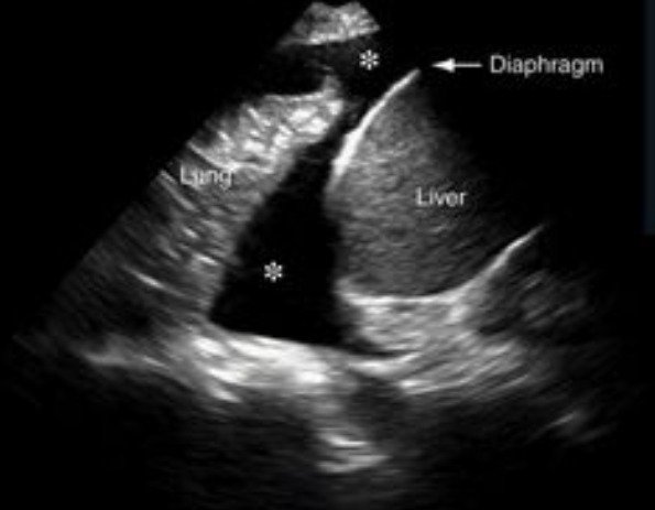
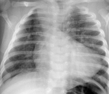


A 4-year-old child with severe pneumonia is mechanically ventilated for hypoxemic respiratory failure. On day 3, oxygen requirement increases (FiO₂ 0.75), PEEP is 8 cmH₂O, and plateau pressures are rising. The child develops tachycardia, increasing ventilator asynchrony, and decreased right-sided breath sounds. Bedside lung ultrasound is performed. The image above is obtained from the right lower thoracic zone using a curvilinear probe.
Quiz Question
• What is the most likely diagnosis based on this ultrasound image, and what is the next best step in management?
Diagnosis
• Large right-sided pleural effusion
Ultrasound Features Visible
• Anechoic (black) fluid collection above the diaphragm
• Compressed, partially collapsed lung (“floating lung” appearance)
• Clear visualization of liver below the diaphragm (confirming right side)
• Diaphragm separating abdominal and thoracic compartments
• This is consistent with a free-flowing pleural effusion, likely parapneumonic in this context.
Physiologic & Clinical Risks (PICU Relevance)
• Reduced functional residual capacity
• Worsening V/Q mismatch
• Increased ventilator pressures
• Risk of progression to:
• Complicated effusion
• Empyema
• Tension hydrothorax (rare but possible under positive pressure ventilation)
• Hemodynamic compromise if large and causing mediastinal shift
Management Strategy
Immediate Priorities
• Assess respiratory mechanics and oxygenation
• Evaluate for septations (suggesting complicated effusion)
• Correlate with inflammatory markers and clinical trajectory
Intervention Indications
• Moderate-to-large effusion with:
• Respiratory compromise
• Failure to improve
• Evidence of loculation
Procedural Management
• Ultrasound-guided thoracentesis (diagnostic and therapeutic)
• Chest tube insertion if:
• Purulent fluid
• pH < 7.2
• Glucose low
• LDH high
• Consider intrapleural fibrinolytics if loculated
• Surgical VATS if failure of drainage
Antibiotics
• Broad-spectrum coverage tailored to:
• Community- vs hospital-acquired infection
• Local resistance patterns
Pearls
• In ventilated children, pleural effusion can significantly worsen lung compliance.
• Ultrasound is more sensitive than chest X-ray for detecting pleural fluid.
• Visualization of the diaphragm and liver confirms correct anatomical orientation.
• Dynamic lung movement within fluid suggests a free-flowing effusion rather than consolidation.