


A 3-month-old infant, weight 4.5 kg, is admitted to the Pediatric Cardiac ICU from the emergency department for worsening cyanosis and episodic desaturations, particularly during agitation and feeding. The infant was born at term, has had poor weight gain, and has not undergone prior cardiac surgery.
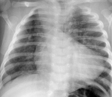
On arrival to the ICU, the child is tachypneic with oxygen saturations fluctuating between 65–75% on supplemental oxygen, heart rate 160/min, and blood pressure appropriate for age. The team obtains a portable chest X-ray, shown above.
No diagnosis has been disclosed to the admitting team yet.
Question
• Based on this chest X-ray, what is the most important abnormality you notice, and what underlying cardiac physiology does it suggest?
Progressive Clues
• Clue 1: Lung fields appear relatively oligemic, without signs of pulmonary edema.
• Clue 2: The cardiac silhouette has an unusual contour, particularly at the cardiac apex and main pulmonary artery segment.
• Clue 3: Oxygen therapy has minimal effect on systemic saturations.
Answer & Explanation
Key Imaging Findings
• On this chest X-ray, the heart demonstrates a characteristic abnormal cardiac silhouette with:
• Uplifted cardiac apex
• Concavity at the level of the main pulmonary artery
• Relatively decreased pulmonary vascular markings
• These findings together create the appearance of a “boot-shaped” heart (coeur en sabot).
Pathophysiologic Correlation
• This radiographic pattern is classic for a cyanotic congenital heart disease with reduced pulmonary blood flow.
• The abnormal silhouette reflects:
• Right ventricular hypertrophy, causing elevation of the cardiac apex
• Hypoplastic or underfilled pulmonary artery segment, leading to the concave left heart border
• Decreased pulmonary blood flow, explaining poor response to oxygen and systemic desaturation
• The cyanosis is driven by right-to-left shunting at the ventricular level, resulting in mixing of deoxygenated blood into systemic circulation.
Differential Diagnosis (Cardiac ICU–Relevant)
• Tetralogy of Fallot
• Boot-shaped heart, pulmonary oligemia
• Often presents with cyanotic spells in early infancy
• Pulmonary atresia with VSD
• More severe pulmonary blood flow reduction
• Often ductal-dependent; may show smaller pulmonary arteries
• Severe pulmonary stenosis with right-to-left shunt
• Similar physiology but usually less classic silhouette
• Tricuspid atresia with decreased pulmonary blood flow
• Typically shows left ventricular dominance on imaging
Management Pearls (Cardiac ICU Focus)
Immediate priorities:
• Ensure adequate oxygenation, but recognize limited response to FiO₂
• Maintain a calm environment to avoid increases in right-to-left shunting
• Optimize preload and systemic vascular resistance
• Continuous cardiorespiratory and saturation monitoring
Red flags:
• Worsening cyanotic spells
• Acidosis or rising lactate
• Sudden drop in pulmonary blood flow or ductal closure (if duct-dependent physiology suspected)
Ventilation considerations:
• Avoid excessive hyperventilation or sudden drops in SVR
• Gentle ventilation strategies if intubation is required
Pearls
• A boot-shaped heart with pulmonary oligemia on CXR is a hallmark of cyanotic congenital heart disease with reduced pulmonary blood flow.
• Chest X-ray remains a powerful physiologic tool in the cardiac ICU when interpreted systematically.
• Cyanosis unresponsive to oxygen should immediately raise concern for intracardiac shunting.
• Image interpretation must always be linked to hemodynamics and physiology, not just pattern recognition.
• Early recognition guides urgent stabilization and cardiology/surgical planning.